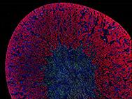
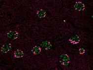
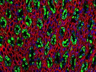
An illuminating cross-section of a kidney A microscopic image scan of a whole mouse kidney. Different microscopic images are digitally stitched together to create this image. The bright fluorescent red structures are the proximal tubuli of the kidney. The nuclei of kidney cells are stained blue. The nuclei of a subpopulation of cells is labeled by a green fluorescent protein.
Day: 16 September 2018
“Bright” podocytes
“Bright” podocytes A microscopic image scan of a whole mouse kidney. Different microscopic images are digitally stitched together to create this image. The bright fluorescent cells are podocytes. This cell type is important for the filtration of the blood. The podocyte cell body is stained in green and the nucleus in pink. Using these fluorescent markers, we can visualize the podocytes and thus the glomeruli (the kidney filters).
A beautiful pattern
A beautiful pattern Fluorescence image of the kidney papilla, which contain tubules that are normally busy processing your urine. The so-called collecting ducts are depicted in green. In red one can see the thin loops of Henle and blood vessels (vasa rectae).