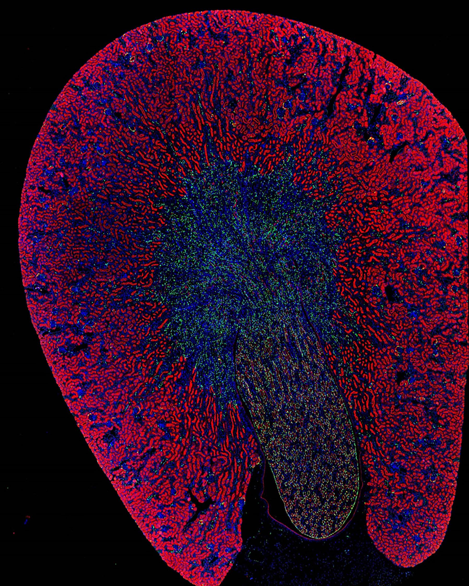
An illuminating cross-section of a kidney
A microscopic image scan of a whole mouse kidney. Different microscopic images are digitally stitched together to create this image. The bright fluorescent red structures are the proximal tubuli of the kidney. The nuclei of kidney cells are stained blue. The nuclei of a subpopulation of cells is labeled by a green fluorescent protein.

