Over membraneuze nefropathie (MN) is de laatste jaren steeds meer bekend geworden. Toch blijven enkele belangrijke vragen onopgehelderd. Celbioloog Bart Smeets van het Radboudumc gaat met een Kolff+ beurs een model ontwikkelen van nierorganoïden om meer kennis te krijgen. Membraneuze nefropathie (MN) is de meest voorkomende oorzaak van het lekken van eiwitten in de urine (nefrotisch syndroom) bij volwassenen. Het is moeilijk te voorspellen hoe de ziekte verloopt en een deel van de mensen heeft uiteindelijk dialyse of een transplantatie nodig om in leven te blijven. Bij ongeveer zeventig procent van de patiënten met MN worden antistoffen gericht tegen PLA2R …
Category: Uncategorized
A huge surface
A huge surface A microscopic image scan of a cross-section of a mouse kidney. Different microscopic images are digitally stitched together to create this image. The bright fluorescent green structures are the brush borders within the kidney. Both kidneys together filter 200 liters of fluid every 24 hours. The filtrate is further processed and concentrated to the 1,5 liter of urine we normally pee throughout the day. The filtrate is processed in specialized tubes (nephrons). A human kidney has about one million nephrons, and the total length of the nephrons in the kidney is about 40 miles! The inside of …
An illuminating cross-section of a kidney
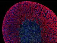
An illuminating cross-section of a kidney A microscopic image scan of a whole mouse kidney. Different microscopic images are digitally stitched together to create this image. The bright fluorescent red structures are the proximal tubuli of the kidney. The nuclei of kidney cells are stained blue. The nuclei of a subpopulation of cells is labeled by a green fluorescent protein.
“Bright” podocytes
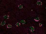
“Bright” podocytes A microscopic image scan of a whole mouse kidney. Different microscopic images are digitally stitched together to create this image. The bright fluorescent cells are podocytes. This cell type is important for the filtration of the blood. The podocyte cell body is stained in green and the nucleus in pink. Using these fluorescent markers, we can visualize the podocytes and thus the glomeruli (the kidney filters).
A beautiful pattern
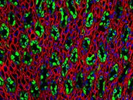
A beautiful pattern Fluorescence image of the kidney papilla, which contain tubules that are normally busy processing your urine. The so-called collecting ducts are depicted in green. In red one can see the thin loops of Henle and blood vessels (vasa rectae).
A cover image
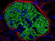
A cover image This is a immunofluorescence staining of a glomerulus of the kidney. Blue: the cell nuclei, red: Basement membrane molecules in Bowman’s capsule and green: synaptopodin, a podocyte specific cytoskeletal protein. This image was used for a cover image
A surprising finding
A surprising finding! These kidney epithelial cells in culture were ignored for several weeks. The cells normally grow as a monolayer on the bottom of the culture flask. Without the necessary care and fresh nutrition, the cells changed their behavior and started to form spheres that were filled with extracellular matrix. This was never shown for this cell type. A finding that was interesting but difficult to reproduce. Nevertheless, a surprising finding.


