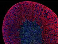
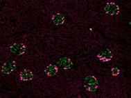
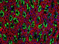
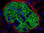
In our pursuit to answer pressing research questions in the kidney pathology, we rely in large part on observations done by microscopic analysis of kidney specimens. The microscope is one of the oldest but also the most advancing tools in the nephropathology.
During the microscopy sessions, we are often blown away by the sheer beauty of the kidney structures. The anatomy of the kidney, which is highly organized, provides the perfect basis for the creation of beautiful images
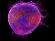
It just felt right to share our microscopical observations from our research. We therefore will share images of our research. These images are in our opinion beautiful aesthetically or “beautiful” because they provide relevant information for our research.
The posts contain images of human and experimental kidney sections but also images of kidney cell cultures.


