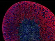
An illuminating cross-section of a kidney A microscopic image scan of a whole mouse kidney. Different microscopic images are digitally stitched together to create this image. The bright fluorescent red structures are the proximal tubuli of the kidney. The nuclei of kidney cells are stained blue. The nuclei of a subpopulation of cells is labeled by a green fluorescent protein.
Author: Kitewebsites
“Bright” podocytes
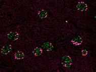
“Bright” podocytes A microscopic image scan of a whole mouse kidney. Different microscopic images are digitally stitched together to create this image. The bright fluorescent cells are podocytes. This cell type is important for the filtration of the blood. The podocyte cell body is stained in green and the nucleus in pink. Using these fluorescent markers, we can visualize the podocytes and thus the glomeruli (the kidney filters).
A beautiful pattern
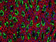
A beautiful pattern Fluorescence image of the kidney papilla, which contain tubules that are normally busy processing your urine. The so-called collecting ducts are depicted in green. In red one can see the thin loops of Henle and blood vessels (vasa rectae).
A cover image
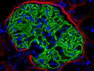
A cover image This is a immunofluorescence staining of a glomerulus of the kidney. Blue: the cell nuclei, red: Basement membrane molecules in Bowman’s capsule and green: synaptopodin, a podocyte specific cytoskeletal protein. This image was used for a cover image
A surprising finding
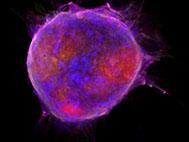
A surprising finding! These kidney epithelial cells in culture were ignored for several weeks. The cells normally grow as a monolayer on the bottom of the culture flask. Without the necessary care and fresh nutrition, the cells changed their behavior and started to form spheres that were filled with extracellular matrix. This was never shown for this cell type. A finding that was interesting but difficult to reproduce. Nevertheless, a surprising finding.
The role of parietal cells in glomerulosclerosis
An interview with researcher Jennifer Eymael about her research into scarring in kidney diseases. This study was published in December 2017 in Kidney International. Interview for NierNieuws (Kidney News) “Niercellen maken littekenvorming zelf erger” (article in Dutch)
Our recent publication in Kidney Int.
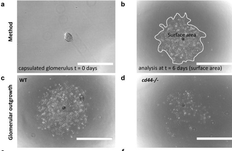
Kidney Int. March 2018, p626–642 CD44 is required for the pathogenesis of experimental crescentic glomerulonephritis and collapsing focal segmental glomerulosclerosis. J. Eymael et al. A key feature of glomerular diseases such as crescentic glomerulonephritis and focal segmental glomerulosclerosis is the activation, migration and proliferation of parietal epithelial cells. CD44-positive activated parietal epithelial cells have been identified in proliferative cellular lesions in glomerular disease. However, it remains unknown whether CD44-positive parietal epithelial cells contribute to the pathogenesis of scarring glomerular diseases. Here, we evaluated this in experimental crescentic glomerulonephritis and the transgenic anti-Thy1.1 model for collapsing focal segmental glomerulosclerosis in CD44-deficient (cd44-/-) …
Older news items
Bart Smeets was selected for the Radboud Hypatia Track. The Radboud Hypatia Track is aimed at excellent researchers who are ready to develop themselves as leaders within their academic fields. Jennifer Eymael won the ‘Best presentation award’ at the “Junge Niere” meeting of the Deutsche Gesellschaft für Nefrologie (DGfN). Jennifer Eymael was awarded with a travel grant for the German annual nephrology congress 2017, Mannheim, Germany. Bart Smeets received the Radboudumc Hypatia Tenure Track grant. Bart Smeets was award winner of the Dr. Werner Jackstädt Research Award 2014. This award is for the promotion and award of outstanding scientists in …
